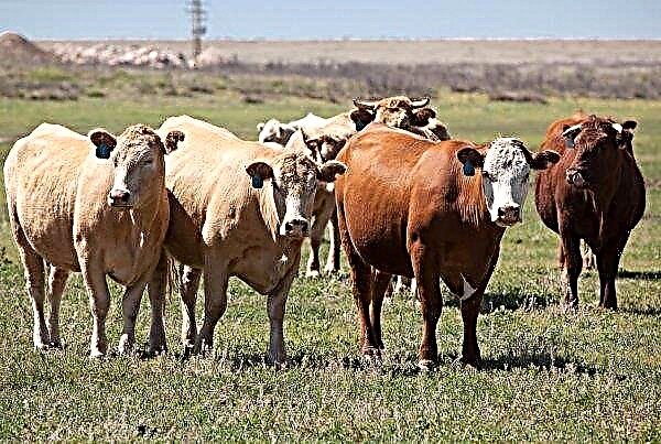
Cattle under inappropriate conditions often infect infectious diseases. The causative agents of these diseases are pathogenic viruses that settle on the mucous membranes of animals. One of the most common infectious diseases in livestock is viral diarrhea. In this article, the causative agent and source of infection with this disease, the course of various forms of diarrhea, as well as possible methods of treatment and the principles of vaccination will be considered.
What is viral diarrhea?
This is a viral infectious disease that is rapidly transmitted from one individual to another. It is expressed in rapid weight loss, diarrhea, accompanied by fever, respiratory disorders and fever.
Important! ATtier maintains its viability at the body of an infected and cured animal for six months and continues to spread through its excretion. Since the virus can be a threat to young animals with weak immunity, it is necessary to keep livestock that was infected with diarrhea in separate cowsheds.
In the absence of treatment, it acquires complications in the form of damage to the joints of the limbs, lameness, inflammation of the cornea of the eyes, stomatitis. Accompanied by a peptic ulcer of the digestive tract.
Economic damage
The damage lies in the massive mortality of cattle in vast territories. In the absence of isolation, it affects not a single farm, but entire regions and regions, so losses are estimated at the state level.
Mortality of infected livestock ranges from 10 to 90%, economic losses are estimated accordingly. When assessing losses, the percentage of mortality, decline in productivity, unborn young growth, and the funds spent on treatment are taken into account.
The causative agent and source of infection
The causative agent of diarrhea is a virus belonging to the genus Pestivirus. It is excreted from the body of an infected animal along with urine, saliva, feces and other physiological secretions. It is related to African swine fever, it affects mainly young animals.
Cattle are infected by contact, through infected feed, water, equipment. The carriers of the virus can be people, birds, insects and rodents.
Symptoms and course of the disease
In total, four forms of the course of this disease are distinguished, which are provoked by the same virus. The forms of infection depend on the physiological state of the animal, its age, susceptibility and the state of the environment in which it is located.
Acute form
Most often develops in young animals - calves up to two months of age. It manifests itself in the form of a strong cough, a sharp increase in body temperature to 41–42 degrees, a depressed state, drowsiness, and apathy. The breathing in infected animals is difficult and shallow, the heart rate exceeds the norm by 1.5 times.
Small ulcerations are observed on the mucous membrane of the nasal passages and oral cavity. Mucous discharge with purulent impurities arbitrarily follows from the nasal passages, there is severe lacrimation and redness of the eyes.
The main symptom is considered to be diarrhea with impurities of blood clots, which lasts for two or more days.
Did you know? For the first time, viral diarrhea was isolated in a separate subspecies in 1946 thanks to the observations of two American farmers named Olafson and Fox. Since then, this disease has become widespread. In the early 1990s, losses from livestock infection were estimated at $ 50 million for every million livestock.
Subacute
It develops in those animals that have developed a certain immunity to this disease. Symptoms in this case are much weaker. Subfebrile body temperature, variable apathy, loss of appetite are observed.
Mucous membranes are affected, but ulcers on them are less pronounced, there is no violation of respiratory function. The cough is shallow, mucous discharge from the nasal passages is insignificant. Lameness is occasionally manifested as a result of inflammatory processes in the joints and short-term diarrhea (up to a day).
Abortive (atypical) form
It proceeds in a half-hidden form, most often occurs in young cattle at the age of four to six months. It manifests itself in the form of a mild, short-term (up to a day) fever, rhinitis, occasionally accompanied by weak lameness and diarrhea without bloody discharge.
Recovery occurs on the fourth day after the onset of symptoms.
Chronic
It is characterized by a weak manifestation of signs of infection, characteristic of animals older than six months of age with formed immunity. There are no inflammatory processes in the oral cavity, there are no lesions of the joints of the limbs.
Interval diarrhea with periods of better health is possible. Such an animal is an active and long-term carrier of the virus, so the chronic form is subject to mandatory treatment.
Diagnostics
Practiced both laboratory and symptomatic. For laboratory tests, samples of the internal organs (lymph nodes, mucous membranes, intestines) of the fallen young animals are taken. Washings and scrapings of the mucous membranes of infected animals are sent to the study, blood sampling is performed for a general analysis. Samples are taken twice - after the onset of symptoms and three weeks after the start of treatment. Symptomatic diagnosis includes examining the mucous membranes of unhealthy livestock, checking its reflexes, and monitoring behavior.
Pathological changes
Changes are analyzed after dissection of the body of a fallen infected animal.
Most often localized in the digestive tract, but is also present on other organs:
- On the mucous membranes of the oral, nasal cavity and esophagus, hyperemia of the vessels, erosive lesions, and surface ulcers of various sizes are observed.
- The esophagus is covered with a gray-brown coating in those places where ulcers occur.
- In the abomasum and scar, point-like vasodilatations, local hemorrhages are found.
- The intestines are filled with fetid masses with the inclusion of blood clots and purulent inclusions.
- The membranes are inflamed, there is swelling and small ulcers covered with mucosal plaque.
- Lymph nodes are noticeably enlarged throughout the body, the liver has a yellow or yellow-orange color.
- The urinary system is inflamed, the kidneys are enlarged, have a flabby soft structure.
Important! Intense diarrhea leads to rapid dehydration of the body, disruption of the water-salt balance, as well as exhaustion. To reduce the death rate, provide the infected livestock with nutritious feed and give them plenty of water.
Treatment
Specific treatments have not been developed. It is possible to strengthen the immunity of an infected livestock by administering blood plasma to previously ill and recovered animals. Relieving the course of the disease is permissible by vaccinating animals with serum from tracheitis or adenovirus disease of cattle. Additional therapeutic measures include the provision of nutritious, easily digestible feeds, plentiful drinking, and the provision of antibiotics to inhibit pathogenic microflora. For intramuscular injections, Levomycetin, Streptomycin, Neomycin, Monomycin and Kanamycin are most often used.
Rinsing of the oral cavity with weak solutions of potassium permanganate and the introduction of interferon isolated from Escherichia coli cultures into the feed are practiced.
Vaccination schedule
Calves fed milk from cows with acquired immunity gain resistance to the causative agent of diarrhea up to a month old. Vaccination is carried out twice every six months with an interval of 30 days of complex inactivated vaccines.
Combovac and Narvak are used against viral diarrhea, rotavirus infection, leptospirosis, parainfluenza and rhinotracheitis.
Other preventive measures
Since methods for effectively combating this disease have not been developed, much attention is paid to preventive measures in farms:
- First of all, infected animals are removed from the herd, their slaughter is organized.
- Disposal of the case is carried out by burning.
- The farm revises the diet of livestock, increases the mass fraction of concentrated feed and vitamin supplements in it.
- Planned disinfecting measures are carried out. Particular attention is paid to cowsheds for pregnant queens and stalls with young animals.
- At the entrance to the production facilities, disinfectant mats are laid that neutralize part of the pathogenic microflora and viruses.
- Once a week, livestock stalls are treated with a misty suspension of iodine solution or acetic acid solution.
 Viral diarrhea is a contagious disease that rapidly affects young cattle. The form of its course depends on the physiological state of the animal, its immunity, as well as the quality and frequency of feeding.
Did you know? Viruses can not be attributed to living organisms, as they are not able to eat and convert food into energy. Everything changes after viruses enter the host organism — they acquire the properties of living units. Viruses begin to multiply, die as a result of the natural selection of stronger units and improve their genetic code. The first virus in the history of mankind was discovered in 1892 - it was a tobacco mosaic virus.
To prevent the occurrence and spread of viral diarrhea, it is necessary to carry out regular preventive measures, and transfer infected animals to quarantine.


